While we live in an age where people are living longer, an important limiting factor on longevity is the ability of the heart to maintain function. Known causes of death for the oldest people on record (over 110 years old) were recorded as heart failure. Heart failure is due to the gradual loss of cardiomyocytes (heart muscle cells) and the increase in scarring of the heart muscle. The process may take place due to low grade inflammation of the muscle, which progresses with age, or injury (such as a heart attack) which may cause a more sudden loss of heart function. Inflammation in the cardiovascular system is common with the aging process, being the result of hypertension, high blood glucose, trigylcerides, or oxidized VDL cholesterol.
Cardiac Aging Characteristics:
Key Conditions of the Aging Heart

Natural support for Cardio Anti-aging
CARDIO VITALITY (Terminalia Arjuna (Rejuna))
YELLOW LONGEVITY (Curcumin, EGCG, Apigenin, Luteolin, Icariin, Carnosic Acid)*
YELLOW NATURALLY (Curcumin, EGCG, Apigenin, Luteolin, Icariin, Carnosic Acid)*
VISION VITALITY MAX (Lutein, Meso Zeaxanthin)
XGEVITY (Glucoraphanin precursor to Sulforaphane)*
*Andrographolide is also included
REFERENCES:
(1) Steenman M, et al. Cardiac aging and heart disease in humans. Biophys Rev. 2017 Apr;
(2) Zhu ZY, et al. Apigenin ameliorates hypertension-induced cardiac hypertrophy and down-regulates cardiac hypoxia inducible factor-lα in rats. Food Funct. 2016 Apr;7
(3) Liu HJ, et al. Apigenin alleviates STZ-induced diabetic cardiomyopathy. Mol Cell Biochem. 2017 Apr
(4) Hu W, et al. Luteolin improves cardiac dysfunction in heart failure rats by regulating sarcoplasmic reticulum Ca2+-ATPase 2a. Sci Rep. 2017 Jan
(5) Oberoi L, et al. The aqueous extract, not organic extracts, of Terminalia arjuna bark exerts cardiotonic effect on adult ventricular myocytes. Phytomedicine. 2011 Feb 15
(6) Parveen A, et al. Terminalia arjuna enhances baroreflex sensitivity and myocardial function in isoproterenol-induced chronic heart failure rats. J Cardiovasc Pharmacol Ther. 2012 Jun
(7) Kaliq F, et al, Improvement in myocardial function by Terminalia arjuna in streptozotocin-induced diabetic rats: possible mechanisms. J Cardiovasc Pharmacol Ther. 2013 Sept.
(8) Kumar S, et al. Proteomic analysis of the protective effects of aqueous bark extract of Terminalia arjuna (Roxb.) on isoproterenol-induced cardiac hypertrophy in rats. J Ethnopharmacol. 2017 Feb 23
(9) Kocak C, et al, Molecular and biochemical evidence on the protective effects of embelin and carnosic acid in isoproterenol-induced acute myocardial injury in rats. Life Sci. 2016 Feb 15
(10) Chung RWS, et al. Lutein exerts anti-inflammatory effects in patients with coronary artery disease. Atherosclerosis. 2017 May 6;
(11) Girandola RN, et al. Effect of E-OJ-01 on Cardiac Conditioning in Young Exercising Adults: A Randomized Controlled Trial. Am J Ther. 2017 May
(12) Qian ZQ, et al. Icariin prevents hypertension-induced cardiomyocyte apoptosis through the mitochondrial apoptotic pathway. Biomed Pharmacother. 2017 Apr.
(13) Gu J, et al. Metallothionein Is Downstream of Nrf2 and Partially Mediates Sulforaphane Prevention of Diabetic Cardiomyopathy. Diabetes. 2017 Feb;
(14) Lin CM, et al. Suppressive effect of epigallocatechin-3-O-gallate on endoglin molecular regulation in myocardial fibrosis in vitro and in vivo. J Cell Mol Med. 2016 Nov;
(15) Tan WS, et al. Is there a future for andrographolide to be an anti-inflammatory drug? Deciphering its major mechanisms of action. Biochem Pharmacol. 2017 Apr 2
(16) Lv FH, et al. Effects of curcumin on the apoptosis of cardiomyocytes and the expression of NF-κB, PPAR-γ and Bcl-2 in rats with myocardial infarction injury. Exp Ther Med. 2016 Dec
(17) Khaliq F, et al. Improvement in myocardial function by Terminalia arjuna in streptozotocin-induced diabetic rats: possible mechanisms. J Cardiovasc Pharmacol Ther, 2013 Sep
(18) Hashemzaei M, et al. Regulation of autophagy by some natural products as a potential therapeutic strategy for cardiovascular disorders. Eur J Pharmacol. 2017 May
(19) Hu J, et al. Luteolin alleviates post-infarction cardiac dysfunction by up-regulating autophagy through Mst1 inhibition. J Cell Mol Med, 2016 Jan
(20) Meghwani H, et al. Beneficial effects of aqueous extract of stem bark of Terminalia arjuna (Roxb.), An ayurvedic drug in experimental pulmonary hypertension. J Ethnopharmocol. 2017 Feb 2
(21) Woo AY, et al. Andrographolide up-regulates cellular-reduced glutathione level and protects cardiomyocytes against hypoxia/reoxygenation injury. J Pharmacol Exp Ther. 2008 Apr
(22) Zhang J, et al. Andrographolide Attenuates LPS-Induced Cardiac Malfunctions Through Inhibition of IκB Phosphorylation and Apoptosis in Mice. Cell Physiol Biochem. 2015
(23) Fernandes RO, et al. Sulforaphane effects on postinfarction cardiac remodeling in rats: modulation of redox-sensitive prosurvival and proapoptotic proteins. J Nutr Biochem. 2016 Aug
New biomarkers have been identified that may help in the early diagnosis and treatment of prostate and urological cancers. Long non-coding RNA (Ribonucleic Acid), a form of RNA not involved in cellular protein encoding, are altered in these cancers.(1,2)
Expression of long non-coding RNA is believed to play a significant role in the initiation and progression of prostate cancer. Sulforaphane has been found to inhibit the expression of non-coding RNA associated with prostate cancer. Researchers conclude that sulforaphane may prevent and suppress prostate cancer by the inhibition of key non-coding forms of RNA. (3)

XGEVITY (contains Glucoraphanin - Sulforaphane precursor)
AIR VITALITY (contains Glucoraphanin - Sulforaphane precursor)
REFERENCES:
(1) Martens-Uzunova ES, et al. Long noncoding RNA in prostate, bladder, and kidney cancer. Eur Urol. 2014 Jun;
(2) Mouraviev B, et al. Clinical prospects of long noncoding RNAs as novel biomarkers and therapeutic targets in prostate cancer. Prostate Cancer Prostatic Dis. 2016 Mar.
(3) Beaver LM, et al. Long noncoding RNAs and sulforaphane: a target for chemoprevention and suppression of prostate cancer. J Nutr Biochem 2017 Apr;
Blue Light from Electronic Technology. Eye Damaging? That is the concern. Everywhere we are constantly exposed to electronic sources of blue light, including smart phones, computer displays, LED and OLED televisions and car lights. While the light emitted from a smartphone is thought to be in the visible spectrum, there is a very high amount of short wave blue light that is also emitted. LED from cars lights, especially at night, may also pose a problem for the retina. Since we are living longer and are exposed continuously to LED lights, there is real danger the retina may be irrevocably harmed.
Natural sunlight (blue light) also causes light-induced damage to the retina, but are less intense than blue light emissions from LED devices. Therefore LED lights significantly increases the potential for toxicity to the retina. (1-4)

BLUE LIGHT DAMAGES RETINA
Photoreceptors (cones and rods) in the retina provide the neuron interface to convert light to images. These photoceptors reside on the outermost parts of the retina, and are nourished and maintained by an underlying layer termed the Retinal Pigment Epithelium (RPE). Photo induced stress directly affects the health of the retina. The blue light is especially damaging, increasing oxidative stress and can lead to cellular death to either the photoreceptors or the Retinal Epithelium.
Age-Related Macular Degeneration (AMD) involves the progressive degradation of the photoreceptors and the RPE. Blue light can damage and cause cellular death of these critical structures. Oxidative stress and inflammation are believed to be key factors in the development of AMD.
NATURAL PROTECTION AGAINST DAMAGING BLUE LIGHT

FURTHER PROTECTION
Bilberry Anthocyanins. Increases Antoxidant Protection of Retina. An experimental model of retinal degeneration, produced by visible-light damage, was ameliorated by the protective antioxidant effects of bilberry anthocyanins. (11)
Sulforaphane. Most potent Nfr2 Activator.
In studies involving the Retinal Pigment Epithelium (RPE), and oxidative stress, sulforaphane was shown to significantly up regulate antioxidant protection of the RPE by activating Nrf2 and HO-1. (12)
Aging Increases Damage from Blue Light in PhotoReceptor cells. Normal protection of the photoreceptor cells and the supporting retiinal epithelium layer, is provided by the cells inherent Nrf2 antioxidant protection against oxidative stress. However, aging is known to deplete the protective Nrf2 response, leaving the retina even more susceptible to damage by oxidative insults such as blue light. Therefore, not only does blue light inherently stress the retina and may cause cellular death, but this response is greatly amplified with retinal aging.(13-14)
VISION VITALITY MAX (Mesozeaxanthin | Zeaxanthin | Lutein | Bilberry)
XGEVITY (Glucoraphinin percursor to Sulforaphane)
REFERENCES:
(1) Coleman S. LED Lights Dangerous on Roadways and Off. 2015 Jan.
(2) Renard G, et al. The dangers of blue light. True story. J Fr Ophtalmol.2016 May.
(3) Jaadane I, et al.Retinal damage induced by commercial light emitting diodes (LEDs) Free Radic Biol Med. 2015 Jul.
(4) Krigel A, et al. Light-induced retinal damage using different light sources, protocols and rat strains reveals LED phototoxicity.Neuroscience, 2016 Dec.
(5) Lima VC, et al. Macular pigment in retinal health and disease. Int J Retina Vitreous. 2016 Aug
(6) Nolan JM, et al. The impact of supplemental macular carotenoids in Alzheimer's disease: a randomized clinical trial. J Alzheimers Dis. 2015
(7) Orthan, et al. Mesozeaxanthin Protects Retina from Oxidative Stress in a Rat Model. Ocul Pharmacol Ther. 2016 Nov
(8) Miyake S, et al. Phase II enzyme induction by a carotenoid, lutein, in a PC12D neuronal cell line. Biochem Biophys Res Commun. 2014 Apr
(9) Zou X, et al. Zeaxanthin induces Nrf2-mediated phase II enzymes in protection of cell death. Cell Death Dis. 2014 May
(10) Kamoshita M, et al. Lutein acts via multiple antioxidant pathways in the photo-stressed retina. Sci Rep. 2016 Jul.
(11) Wang Y, et al. Retinoprotective Effects of Bilberry Anthocyanins via Antioxidant, Anti-Inflammatory, and Anti-Apoptotic Mechanisms in a Visible Light-Induced Retinal Degeneration Model in Pigmented Rabbits. Molecules. 2015 Dec
(12) Ye L, et al, Sulforaphane enhances the ability of human retinal pigment epithelial cell against oxidative stress, and its effect on gene expression profile evaluated by microarray analysis. Oxid Med Cell Longev, 2013
(13) Sachdeva MM, et al. Nrf2 signaling is impaired in the aging RPE given an oxidative insult. Exp Eye Res. 2014 Feb;
(14) Chen WJ, et al. Nrf2 protects photoreceptor cells from photo-oxidative stress induced by blue light. Exp Eye Res. 2016 Dec
MITOPHAGY AND LONGEVITY
NRF2 - THE ROLE IN MITOPHAGY AND LONGEVITY Nrf2 is a latent protein in the cell, which upon activation, regulates the activation of genes which produce antioxidant proteins for cellular protection, reduction of inflammation and reduction of mitochondrial toxins (via glutathione induction).
REFERENCES:
(1) Palikaras K, et al. Mitophagy: In sickness and in health. Mol Cell Oncol. 2015 Jun.
(2) Palikaras K, et al. Coupling mitogenesis and mitophagy for longevity. Autophagy. 2015.
(3) LaPierre L, et al. Transcriptional and epigenetic regulation of autophagy in aging. Autophagy. 2015 Jun
(4) Greco T, et al. Sulforaphane Inhibits Mitochondrial Permeability Transition and Oxidative Stress. Free Radic Biol Med, 2012 Dec
(5) Holstrom Kira, et al. The multifaceted role of Nrf2 in mitochondrial function. Curr Opin Toxicol. 2016 Dec
(6) Wang K, et al. Redox homeostasis: the linchpin in stem cell self-renewal and differentiation. Cell Death Dis. 2013 Mar
(7) Russo M, et al. Nrf2 targeting by sulforaphane: a potential therapy for cancer treatment. Crit Rev Food Sci Nutr. 2016 Dec
(8) O'Mealey GB, et al. Sulforaphane is a Nrf2-independent inhibitor of mitochondrial fission. Redox Biol. 2016 Nov
Air pollution is a pervasive and often ignored factor in the aging process. Airborne ultra-fine (nanoparticles) pollution particles are microscopic and pose special dangers to health. Researchers agree that ultra-fine air pollution particles are a major threat in negative health consequences in humans. (1) While being very detrimental to the lungs, long-term exposure to ultra-fine particles also damages the cardiovascular system, contributes to atherosclerosis plaque formation and may lead to degenerative brain diseases - including Alzheimer's disease. These very small particles are able to travel deep into lung tissue and into the blood stream. As a result, there are significant increases in systemic oxidative stress, inflammation with increased DNA damage and mutagenicity. (2-6) Telomere length, which is an indicator of biological aging, becomes shorter in people living in air pollution areas. Shorter telomeres is associated with accelerated aging.(7-8)

Neuroinflammation. Especially alarming is the effect of ultra-fine particles on the brain. Airborne nano-sized particulate matter migrates through the lungs into the bloodstream and eventually into the brain. Longtime exposure to ultra-fine air pollutants leads to chronic brain inflammation which leads to neurodegenerative diseases. Epidemiological research has linked ultra-fine air particulate matter as a significant environmental factor involved in Alzheimer's Disease and Parkinson's Disease.(2)
Ultra-fine pollutant particles are produced by high heat sources including automobiles, trucks, airplane exhaust and factories and play a significant role in health and longevity. Urban areas, transit routes (highways and roads) and airports are major sources of pollutants but the suburbs are not immune. Such pollutants are so small that most people are unaware that they are inhaling toxic pollutants. Airborne fine particulate matter is a major component of air pollution.
Sources of ultra-fine particle pollutants:
(1) City / Urban Areas Particulate Matter. Urban areas create a confluence of major activity which generate ultra-fine air pollution. In studies involving six China urban areas, ultra-fine particulate matter (PM), including polycyclic aromatic hydrocarbon (PAH), were analyzed. The particulate matter was determined as having cytotoxicty to the bronchial cells of the lungs.(9) Substantial sources of outdoor PAHs in particulate pollution include cars and trucks.
(2) Highways / roadways. high volumes of traffic on highways are significant sources of ultra-fine particulate matter and PAH which is then distributed to surrounding areas and runoff into water ways. People living close to major roadways have increased health risks due increased air pollution exposure. Road paving and asphalt manufacturing are also major sources.
(3) Indoor sources. Burning of biofuels including coal and wood create dangerous levels of emissions. Also emissions from cigarette smoke.
(4) Diesel exhaust - classified as a group 1 carcinogen. Shown to cause lung and possibly bladder cancer.Contains nitric oxide and fine particulate matter.
(5) Airports / Aircraft turbine engines - these aircraft engines produce significant levels of hazardous ultra-fine air pollutants (including benzene and formaldehyde) at both in flight and ground levels. In fact, air quality impacts areas extend far from the airports. The LA times reports that communities as far as 10 miles from LAX airport are exposed to unsafe levels of air pollutants from aircraft engine exhaust.(10)
Natural Support:
SULFORAPHANE - Sulforaphane is a potent activator of phase 2 detoxification in the body. In a study in high air pollution areas in China, sulforaphane beverages were shown to signifcantly increase metabolism and excretion of hazardous air pollutants benzene and acrolein in participants.(11) Further studies indicate that through activation of Nrf2, sulforaphane enhances protection against airborne toxin carcinogens and carcinogenesis. (12) Sulforaphane also protects against oxidant effects of diesel emission by invoking an over expression of antioxidant enzymes.(13)
N-ACETYL-CYSTEINE - Offers protection against air pollution induced inflammation. Includes specific mitigation against formaldehyde induced lung damage. Formaldehyde is a major emission toxin from airplane exhaust.(14)
XGEVITY (Glucoraphanin / Sulforaphane and N-Acetyl-Cysteine)
AIR VITALITY (Glucoraphanin / Sulforaphane and N-Acetyl-Cysteine)
REFERENCES:
(1) Chen R, et al. Beyond PM2.5: The role of ultrafine particles on adverse health effects of air pollution. Biochim Biophys Acta. 2016 Dec
(2) Heusinkveld HJ, et al. Neurodegenerative and neurological disorders by small inhaled particles. Neurotoxicology. 2016 Sep
(3) Valavanidis A, et al. Airborne particulate matter and human health: toxicological assessment and importance of size and composition of particles for oxidative damage and carcinogenic mechanisms. Environ Sci Health C Environ Carcinog Ecotoxicol Rev. 2008 Oct-DecBottom of Form
(4) Pope CA, et al. Exposure to Fine Particulate Air Pollution Is Associated with Endothelial Injury and Systemic Inflammation. Circ Res. 2016 Oct 25
(5) Bai Y, et al. Fine particulate matter air pollution and atherosclerosis: Mechanistic insights. Biochim Biophys Acta. 2016 Dec
(6) Risom L, et al. Oxidative stress-induced DNA damage by particulate air pollution. Mutat Res. 2005 Dec 30
(7) Martens Ds, et al. Air Pollution Stress and the Aging Phenotype: The Telomere Connection. Curr Environ Health Rep. 2016 Sep
(8) Ward-Caviness CK, et al. Long-term exposure to air pollution is associated with biological aging. Oncotarget 2016 Oct 25
(9) Yang L, et al. Pro-inflammatory response and oxidative stress induced by specific components in ambient particulate matter in human bronchial epithelial cells. Environ Toxicol. 2016 Aug
(10) Ridgeway L. New concerns raised about air pollution at LAX. May 30, 2014
(11) Egnar PA, et al. Rapid and sustainable detoxication of airborne pollutants by broccoli sprout beverage: results of a randomized clinical trial in China.
(12) Kensler TW, et al. Keap1-nrf2 signaling: a target for cancer prevention by sulforaphane. Top Curr Chem. 2013.
(13) Wan J, et al. Antioxidant enzyme induction: a new protective approach against the adverse effects of diesel exhaust particles. Inhal Toxicol 2007.
(14) Wang M, et al. N-acetylcysteine: A promising drug against formaldehyde-induced damage in lung epithelial cells. Med Hypotheses. 2014 Nov
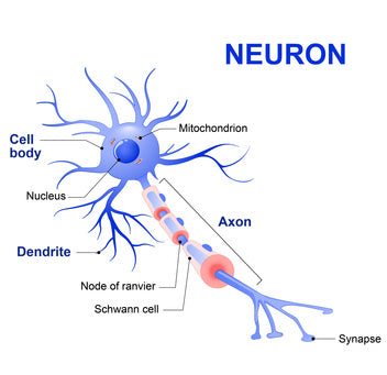
Aging of the brain involves the loss of neurons (hippocapmus shrinkage), loss of synapse integrity between neurons, build-up of toxic amyloid proteins, neuron tangles, defects in blood flow and chronic inflammation. Under normal age progression, these events do not happen over night and may take years before impairments in cognition become noticed.
Ultimate anti-aging strategies for the brain and memory should target the progressive decline of the brain and promote reversal and recovery of some cognition impairments.
Emerging research in the study of herbal ingredients show their tremendous potential use in mitigating the decline in brain function with age.
Herbs and Extracts
- Andrographolide - As an extract from Andrographis Paniculata. Lab research has shown stimulation of neurogenesis in the hippocampus by andorgrapholide. Specifically " increased cell proliferation and the density of immature neurons in the dentate gyrus." (1) The dentate gyrus is an area of the hippocampus involved in memory formation.
- Centella asiatica - Acts as a potent memory enhancer, via increasing hippocampus neurogenesis and support for brain tissue regeneration. (2)
- Baicalin (3)
- Panax Ginseng (4)
- Curcumin (5)
- Epimedium (Icariin) (6)
- Apigenin (7)
Herbs and Extracts
- Centella asiatica - Improves structural integrity of axons / myelination and proliferation of dendritic branching and length. Such improvements have been shown to enhance learning and improve memory. (8) Centella asiatica also has been shown to improve learning and memory in normal lab mice.
- Luteolin - Baicalin - promote neuronal survival and neuron differentiation through the outgrowth of neurites (axons and dendrites) from the neuron.(9.10)
- Rosemary (Carnosic Acid) - Strongly promotes neurite outgrowth as a function of powerful Nrf2 activity. Suppressed Nrf2 activation suppresses neuron differentiation.(11)
Herbs and Extracts
- Andrographolide - Impairment of synaptic function between neurons plays a significant role in the loss of cognitive function. This is seen in the progression of AD. In research animals with AD-like cognitive disease, the treatment of andrographolide over a 3 month span imporved synaptic function and protected important synaptic proteins.
- Furthermore, andrographolide has been shown to reduce inflammation in the brain and levels of pathological tau protein and beta amyloid in animal models.(12)
- Andrographolide reduces inflammation and dysfunction of the cerebral endothelial cells, which may affect vascular flow to the brain.(13)
- Centella asiactica - in senescence-accelerated lab mice, which had accelerated aging of the brain, administration of centella asiatica significantly improved synaptic plasticity and reduced beta amyloid build-up. Such treated mice showed significant benefits in memory and learning. (14)
REFERENCES:
NEUROGENESIS
(1) Varela-Nallar L, et al. Andrographolide Stimulates Neurogenesis in the Adult Hippocampus. Neural Plast, 2015.
(2) Sirichoat A, et al. Effects of Asiatic Acid on Spatial Working Memory and Cell Proliferation in the Adult Rat Hippocampus. Nutrients. 2015 Oct 5
(3) Zhang K, et al. Baicalin promotes hippocampal neurogenesis via SGK1- and FKBP5-mediated glucocorticoid receptor phosphorylation in a neuroendocrine mouse model of anxiety/depression. Sci Rep. 2016 Aug 9
(4) Jiang B, et al. Antidepressant-like effects of ginsenoside Rg1 are due to activation of the BDNF signalling pathway and neurogenesis in the hippocampus. Br J Pharmacol. 2012 Jul;
(5) Pluta R, et al. Neurogenesis and neuroprotection in postischemic brain neurodegeneration with Alzheimer phenotype: is there a role for curcumin? Folia Neuropathol. 2015
(6) Li F, et al. Icariin decreases both APP and Aβ levels and increases neurogenesis in the brain of Tg2576 mice. Neuroscience. 2015 Sep 24
(7) Taupin P. Apigenin and related compounds stimulate adult neurogenesis. Mars, Inc., the Salk Institute for Biological Studies: WO2008147483. Expert Opin Ther Pat. 2009 Apr
BRAIN TISSUE REGENERATION
(8) Yogeswarin L, et al. Recent Updates in Neuroprotective and Neuroregenerative Potential of Centella asiatica. Malays J Med Sci 2016 Jan.
(9) Chen PY, et al. Up-Regulation of miR-34a Expression in Response to the Luteolin-Induced Neurite Outgrowth of PC12 Cells. J Agric Food Chem. 2015 Apr
(10) Li M, et al. Neuronal differentiation of C17.2 neural stem cells induced by a natural flavonoid, baicalin. Chembiochem. 2011 Feb 11;
(11) Kosaka K, et al. Role of Nrf2 and p62/ZIP in the neurite outgrowth by carnosic acid in PC12h cells. J Biochem. 2010 Jan;
RECOVERING COGNITIVE DYSFUNCTION
(12) Rivera DS, et al. Andrographolide recovers cognitive impairment in a natural model of Alzheimer's disease (Octodon degus). Neurobiol Aging. 2016 Jul 5
(13) Chang CC, et al. Andrographolide, a Novel NF-κB Inhibitor, Inhibits Vascular Smooth Muscle Cell Proliferation and Cerebral Endothelial Cell Inflammation. Acta Cardiol Sin. 2014 Jul;
(14) Xing L, et al. Beneficial effects of asiaticoside on cognitive deficits in senescence-accelerated mice. Fitoterapia. 2013 Jun.
NEUROPROTECTION - AMYLOID TOXICITY AND INFLAMMATION
(15) Gray NE, et al. Centella asiatica Attenuates Amyloid-β-Induced Oxidative Stress and Mitochondrial Dysfunction. J Alzheimers Dis. 2015
(16) Zhang L, et al. Icariin reduces α-synuclein over-expression by promoting α-synuclein degradation. Age (Dondr.) 2015 Aug
(17) Chen YJ, et al. Neuroprotective Effects of Icariin on Brain Metabolism, Mitochondrial Functions, and Cognition in Triple-Transgenic Alzheimer's Disease Mice. CNS Neurosci Ther, 2016 Jan
(18) Dirscherl K, et al. Luteolin triggers global changes in the microglial transcriptome leading to a unique anti-inflammatory and neuroprotective phenotype. J Neuroinflammation 2010 Jan
(19) Rezai-Zedeh K, et al. Apigenin and luteolin modulate microglial activation via inhibition of STAT1-induced CD40 expression. J Neuroinflammation. 2008 Sep
(20) Chen C, et al. Baicalin attenuates alzheimer-like pathological changes and memory deficits induced by amyloid β1-42 protein. Metab Brain Dis. 2015 Apr
(21) Song F, et al. Schizandrin A Inhibits Microglia-Mediated Neuroninflammation through Inhibiting TRAF6-NF-κB and Jak2-Stat3 Signaling Pathways. PLoS One. 2016 Feb 26;
(22) Habtemariam S. The Therapeutic Potential of Rosemary (Rosmarinus officinalis) Diterpenes for Alzheimer's Disease. Evid Based Complement Alternat Med, 2016
REVERSES BRAIN INSULIN RESISTANCE IN BRAIN NEURONS
(23) Feng HL, et al. Curcumin ameliorates insulin signalling pathway in brain of Alzheimer's disease transgenic mice. Int J Immunopathol. 2016 Jul 27
(24) Angeloni C, et al. Neuroprotective effect of sulforaphane against methylglyoxal cytotoxicity. Chem Res Toxicol, 2015 Jun 15
Air pollutants are harmful and affect everyone, but may be additionally detrimental for people who exercise at higher levels (with higher inhalation rates) both outdoors and indoors. Since runners, bikers, aerobics class participants and other fitness buffs, require more oxygen and deeper inhalation. they typically inhale higher level of pollutants. For example, a fitness center study comparing the inhalation of pollutants of indoor aerobics class vs. less intense activity, showed 2x's the amount of pollutants inhaled by the higher intensity aerobics class. (1) Moreover, the air pollutants tend to become trapped deeper into the lung tissue. Furthermore, air pollutants are most often not apparent and the fitness ethusiast is unlikely to be aware of the inhalation hazard.
Air pollutants have been associated with respiratory mortality (3), development of atherosclerosis (2), diabetes (5) and neuropatholgies (via increased neuro inflammation) (4). While doctors support the need for exercise, many will caution about exercising outdoors, especially where air pollution levels may be higher.
SOURCES OF AIR POLLUTION:
SULFORAPHANE DETOXIFICATION
Sulforaphane is a powerful phase 2 detoxifier, which enables the detoxfication and excretion of harmful pollutants and toxins. In a clinical trial in china, glucoraphain in broccoli sprout (beverage), which contains the powerful precursor of sulforaphane, was shown to effectively detoxify several major air pollutants.(6) Compared to placebo, the broccoli sprout beverage showed "rapid and sustained increases in the levels of excretion of the glutathione-derived conjugates of benzene (61%), acrolein (23%)". Another study, also showed increased excretion of the air pollutant crotonaldehyde.(7)
Effective results showed detoxification of airborne pollutants from study participants.(6,7) Sulforaphane may also have an inhibitory effect against cancer, including lung cancer, via activation of Nrf2 antioxidant pathway. (10)
Diesel Exhaust Fine Particles - The fine particles form diesel exhaust are considered a significant health hazard which impacts especially outdoor exercise. At high cardiac output, diesel increased pulmonary vasoconstriction (8) and increased oxidative stress and allergic inflammatory response.(9) Sulforaphane induces phase II enzymes which can block the oxidant and allergic inflammation harmful effect of the diesel exhaust particles. (9)
XGEVITY (Sulforaphane precursor Glucoraphanin)
REFERENCES:
(1) Ramos CA, et al. Estimating the inhaled dose of pollutants during indoor physical activity. Sci Total Environ. 2015 Sep 15
(2) Bai Y, et al. Fine particulate matter air pollution and atherosclerosis: Mechanistic insights. Biochim Biophys Acta. 2016 May 6.
(3) Brunekreef B, et al. Effects of long-term exposure to traffic-related air pollution on respiratory and cardiovascular mortality in the Netherlands: the NLCS-AIR study. Res Rep Health Eff Inst. 2009 Mar
(4) Jørgensen JT, et al. Long-term exposure to ambient air pollution and incidence of brain tumours: The Danish Nurse Cohort. Neurotoxicology. 2016 Jun 2
(5) Hansen AB, et al. Long-term exposure to fine particulate matter and incidence of diabetes in the Danish Nurse Cohort. Environ Intl 2016 May;
(6) Egner PA, et al. Rapid and sustainable detoxication of airborne pollutants by broccoli sprout beverage: results of a randomized clinical trial in China. Cancer Prev Res (Phila). 2014 Aug
(7) Kensler TW, et al. Modulation of the metabolism of airborne pollutants by glucoraphanin-rich and sulforaphane-rich broccoli sprout beverages in Qidong, China. Carcinogenesis. 2012 Jan
(8) Wauters A, et al. At high cardiac output, diesel exhaust exposure increases pulmonary vascular resistance and decreases distensibility of pulmonary resistive vessels.
(9) Wan J, et al. Antioxidant enzyme induction: a new protective approach against the adverse effects of diesel exhaust particles. Inhal Toxicol. 2007
(10) Yang L, et al. Frugal chemoprevention: targeting Nrf2 with foods rich in sulforaphane. Semin Oncol. 2016 Feb
Subscribe to our newsletter and always be the first to hear about what is happening.
© 2024 Geres Dengle®